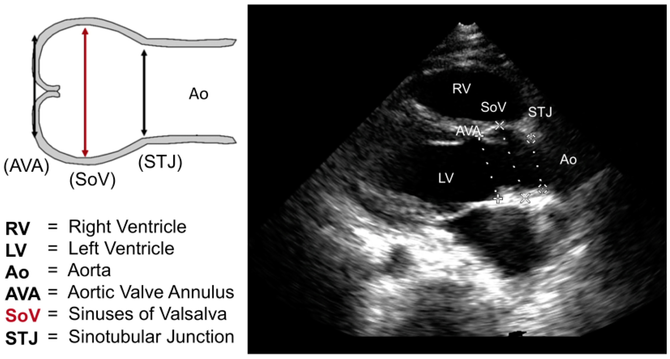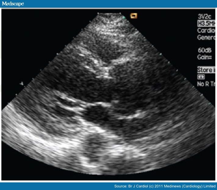
Marfan syndrome is often diagnosed in patients who are tall due to their long limbs, arachnodactyly, pectus deformities, and scoliosis. An aortic root aneurysm, if left untreated, can cause an enlarged pulse pressure or diastolic murmur, both of which can lead to heart failure. Bicuspid valves, as well as those with Marfan’s syndrome, can cause a rapid expansion of the aorta. In 15% of cases, Takayasu’s arteritis causes aorticular dilation, which is often obstructive. When an ascending aortic wall is infected by a true primary bacterial infection, there is a very small chance of an aortic aneurysm developing, either from an episode of bacterial endocarditis or from an infection of an existing aortic wall. In contrast to the supravalvular aneurysm that can be treated with a simple supracoronary tube graft, the aortic rootneurysm requires the aortIC valve to be spared or replaced. It can be divided into two distinct categories based on its prevalence, but it accounts for half of all thoracic aneurysms. If the aorta thickens without surgical intervention, the risk of ascending aortic dissection or aortic rupture increases. Treatment options include open surgery to repair the aneurysm or endovascular surgery to place a stent in the aorta to help keep it open. If your aortic aneurysm is large, growing quickly, or causing symptoms, your doctor may recommend treatment to prevent it from rupturing. If you have an aortic aneurysm, your doctor will monitor it closely with periodic imaging tests.

Most aortic aneurysms do not cause symptoms and are found during a routine medical exam or imaging test for another condition.

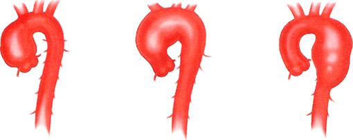
Aortic aneurysms can rupture, or tear open, causing life-threatening bleeding. This is the portion from the Aortic Arch to the Diaphragm, the muscle that separates the chest from the abdomen.īelow CT images of a descending thoracic aorta, left before treatment and on the right following procedure.An aortic aneurysm is when the aorta, the main blood vessel that carries blood from the heart to the rest of the body, enlarges or ballooning. Note the close proximity of head vessels to the dilated aortic aneurysm:ĭescending Thoracic Aortic Aneurysm – straight part of aorta in the back of the chest carrying blood to the lungs, spinal cord and abdomen. Intraoperative photograph of a large ascending aortic aneurysm.Īortic Arch Aneurysm – part of the aorta that supplies blood to the head and neck.ĬT images of arch aneurysms. Treatment for Ascending Aortic Aneurysm – Note the contrast dye that is “leaking” back into the heart.Īscending Aortic Aneurysm – curved part of the aorta in the front of the chest above the coronary arteries and before the arch. Relatives of patients with thoracic aortic aneurysms should undergo an elective screening test such as echocardiography, CT or MRI.Īneurysms can occur in one or more of the various portions of the thoracic aorta:Īortic Root Aneurysm – portion of the aorta closest to the heart and involves the arteries that supply blood and oxygen to the heart.Īngiographic picture of large aortic root with a severely leaking aortic valve. Thus, when diagnosed they should be followed by a specialized team. However, in most cases thoracic aortic aneurysms tend not to cause symptoms until it is too late.

As the aorta gets larger, there is a risk that the wall of the aorta can tear or burst.Īs the aneurysm grows there can be symptoms of shortness of breath, especially if the associated valve is leaking. The aorta carries the entire blood flow out of the heart and supplies blood and oxygen to the brain, chest and abdominal organs.

Thoracic Aortic Aneurysms are a dilation or growth of the aorta due to a weakening in the wall of the artery.


 0 kommentar(er)
